Home
/ Animal Cell Diagram Edgenuity : Education Chart Biology Animal Cell Diagram Stock Vector ... / The result is two centrosomes, each with its own pair of centrioles.
Animal Cell Diagram Edgenuity : Education Chart Biology Animal Cell Diagram Stock Vector ... / The result is two centrosomes, each with its own pair of centrioles.
Animal Cell Diagram Edgenuity : Education Chart Biology Animal Cell Diagram Stock Vector ... / The result is two centrosomes, each with its own pair of centrioles.. Diagram of animal cell anatomy illustration. A clear design animal cell diagram template from edraw is waiting for you in the free download version. I spelt it wrong in the diagram, sorry). Use it for any kinds of science coursework or group discussions. A strong, supporting layer around the in plants, algae, and some bacteria.
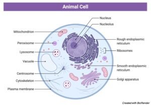
Education chart of biology for animal and plant cell diagram. Cells consist of cytoplasm enclosed within a membrane, which contains many biomolecules such as proteins and nucleic acids.2 most plant and animal cells are only visible under a light microscope, with dimensions between 1 and 100 micrometres.3 detailed diagram of lipid bilayer cell membrane. Animal cells are the basic unit of life in organisms of the kingdom animalia. Water, waste products, food, and other cellular material. A strong, supporting layer around the in plants, algae, and some bacteria.
Animal Cell: Definition, Diagram, Structure, and Function from researchtweet.com Some students may be able to identify some of the structures. The diagram, like the one above, will include labels of the major parts of an animal cell including the cell membrane, nucleus, ribosomes, mitochondria, vesicles, and cytosol. Cells consist of cytoplasm enclosed within a membrane, which contains many biomolecules such as proteins and nucleic acids.2 most plant and animal cells are only visible under a light microscope, with dimensions between 1 and 100 micrometres.3 detailed diagram of lipid bilayer cell membrane. Diagram of animal cell anatomy illustration. In 5 minutes.this video is specifically for beginners.continue. A comparison of plant and animal cells using labelled diagrams and descriptive explanations. Then, on the next page, define and describe the function of each organelle present. There are three microtubules in each group.
Cell is a tiny structure and functional unit of a living organism containing various parts known as organelles.
Animal cell anatomy diagram structure with all parts nucleus smo. All cells are produced from other cells. Cellular water levels biological vector illustration diagram with animal and plant cell. Cytoplasm, ribosomes, rough endoplasmic reticulum; · an animal cell diagram is a great way to learn and understand the many functions of an animal cell. Each centriole is a ring of nine groups of fused microtubules. Cells consist of cytoplasm enclosed within a membrane, which contains many biomolecules such as proteins and nucleic acids.2 most plant and animal cells are only visible under a light microscope, with dimensions between 1 and 100 micrometres.3 detailed diagram of lipid bilayer cell membrane. Cells are the basic units of structure and function in living things. A system of flattened membranes called cisternae (mainpoint: All organisms are made up of cells (or in some cases, a single cell). I spelt it wrong in the diagram, sorry). Unlike the eukaryotic cells of plants and fungi, animal cells do not have a cell wall. Have cell walls and chloroplasts in contrast to animal cells which have no cell wall or chloroplasts.
I spelt it wrong in the diagram, sorry). An animal cell is the smallest unit that makes up the varied tissues of animal species. Vector diagram for your design, educational, medical, biological and science use. Animal cells are the basic unit of life in organisms of the kingdom animalia. Under the microscope, an animal cell shows many different parts called organelles, that work together to keep the cell functional.
Plant Cells Vs. Animal Cells (With Diagrams) | Owlcation from usercontent2.hubstatic.com Education chart of biology for animal and plant cell diagram. In 5 minutes.this video is specifically for beginners.continue. One of the most problematic duties that health and wellbeing experts face throughout their interplay with patients helps them recognise the issues and how to encourage them about the analysis and therapy available. These parts are called subcellular structures. Furthermore, experts often refer to them as the building blocks of life. below is a diagram of a generalized animal cell. Animal cell anatomy diagram structure with all parts nucleus smo. All animal cells are made up of various different parts. The largest organelle within the cell.
Each centriole is a ring of nine groups of fused microtubules.
In 5 minutes.this video is specifically for beginners.continue. Education chart of biology for animal and plant cell diagram. Have cell walls and chloroplasts in contrast to animal cells which have no cell wall or chloroplasts. There are hundreds of cell types in a developed organism, which are specific to their location and function. Animal cell anatomy diagram structure with all parts nucleus smo. All cells are produced from other cells. Cells consist of cytoplasm enclosed within a membrane, which contains many biomolecules such as proteins and nucleic acids.2 most plant and animal cells are only visible under a light microscope, with dimensions between 1 and 100 micrometres.3 detailed diagram of lipid bilayer cell membrane. Then, on the next page, define and describe the function of each organelle present. Animal cells are packed with amazingly specialized structures. As we all know, the cell is certainly the smallest unit of life. Diagram of animal cell anatomy illustration. Under the microscope, an animal cell shows many different parts called organelles, that work together to keep the cell functional. A clear design animal cell diagram template from edraw is waiting for you in the free download version.
As we all know, the cell is certainly the smallest unit of life. The red blood cells make up the blood, while the nerve cells make up the nervous system tissues. Water, waste products, food, and other cellular material. Instead, multicellular animals have other structures that provide support to their tissues. This is an online quiz called animal cell diagram.
30 Animal Cell Model Diagram And How To Understand Them ... from i.pinimg.com The cell is the basic unit of life. Smooth endoplasmic reticulum, mitochondria, golgi bodies, lysosomes. There are three microtubules in each group. The diagram, like the one above, will include labels of the major parts of an animal cell including the cell membrane, nucleus, ribosomes, mitochondria, vesicles, and cytosol. The result is two centrosomes, each with its own pair of centrioles. An animal cell is the smallest unit that makes up the varied tissues of animal species. Unlike the eukaryotic cells of plants and fungi, animal cells do not have a cell wall. In 5 minutes.this video is specifically for beginners.continue.
During animal cell division, the centrioles replicate (make new copies) and the centrosome divides.
Unlike the eukaryotic cells of plants and fungi, animal cells do not have a cell wall. Animal cell anatomy diagram premium vector. There are hundreds of cell types in a developed organism, which are specific to their location and function. Then, on the next page, define and describe the function of each organelle present. The cell is the basic unit of life. Have cell walls and chloroplasts in contrast to animal cells which have no cell wall or chloroplasts. Table of contents definition explanation diagram structure types conclusion let us have a detailed overview of the animal cell, its types, diagram and structure. A system of flattened membranes called cisternae (mainpoint: Check this diagram and learn m. Show the animal cell diagram to the student(s) and ask what they think it is. There is a printable worksheet available for download here so you can take the quiz with pen and paper. Each centriole is a ring of nine groups of fused microtubules. Cells consist of cytoplasm enclosed within a membrane, which contains many biomolecules such as proteins and nucleic acids.2 most plant and animal cells are only visible under a light microscope, with dimensions between 1 and 100 micrometres.3 detailed diagram of lipid bilayer cell membrane.
Share :
Post a Comment
for "Animal Cell Diagram Edgenuity : Education Chart Biology Animal Cell Diagram Stock Vector ... / The result is two centrosomes, each with its own pair of centrioles."


Post a Comment for "Animal Cell Diagram Edgenuity : Education Chart Biology Animal Cell Diagram Stock Vector ... / The result is two centrosomes, each with its own pair of centrioles."