Animal Cell Diagram With Organelles / Labeling Organelles Practice Animal Cell Diagram Quizlet : The core nucleus, endoplasmic reticulum, golgi apparatus, lysosomes, ribosomes, mitochondria, centriole.. The following points highlight the ten main types of cell organelles present in the cell. Animal cell anatomy diagram structure with all parts nucleus smo. In truth, there are still features of plant and animal cells we're only lately discovering. He explains each organelle's function including the nucleus, nucleolus, nuclear envelope, nuclear. A system of flattened membranes called cisternae (mainpoint:
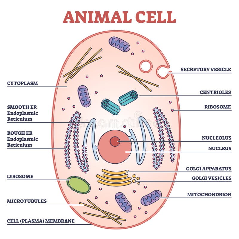
Though this animal cell diagram is not representative of any one particular type of cell, it provides insight into the primary organelles and the intricate internal structure of most animal cells. Cell organelles types (with diagram). Printable animal cell diagram to help you learn the organelles in an animal cell in preparation for your test or quiz. Cytoplasm, ribosomes, rough endoplasmic reticulum; Smooth endoplasmic reticulum, mitochondria, golgi bodies, lysosomes.
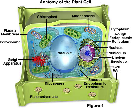
Animal Cell Stock Illustrations 9 557 Animal Cell Stock Illustrations Vectors Clipart Dreamstime from thumbs.dreamstime.com To check if you have understood the cell parts, draw a. Animal cells are eukaryotic in nature, possessing a nucleus and organelles that carry out the different functions the cell must do to thrive and reproduce. In plants and some algae, organelles known as chloroplasts serve as the site of photosynthesis. An animal cell diagram is a great way to learn and understand the many functions of an animal cell. A system of flattened membranes called cisternae (mainpoint: Chloroplasts contain a pigment known as chlorophyll, which captures the sun's energy to transform water and. Cell organelles types (with diagram). Unlike the eukaryotic cells of plants and fungi, animal cells do not have a cell wall.
An animal cell diagram is a great way to learn and understand the many functions of an animal cell.
A system of flattened membranes called cisternae (mainpoint: Education chart of biology for animal and plant cell diagram. There are many types of organelles, particularly in eukaryotic cells. Printable animal cell diagram to help you learn the organelles in an animal cell in preparation for your test or quiz. But at the same time it is interpretive. A micrograph of animal cells, showing the nucleus (stained dark red) of each cell. The animal cell has 13 different types of organelles¹ with specialized functions. All animal cells contain organelles. Though this animal cell diagram is not representative of any one particular type of cell, it provides insight into the primary organelles and the intricate internal structure of most animal cells. The largest organelle within the cell. The animal cell and plant cell diagrams are easily colorable, allowing students to differentiate the different parts of the cell quickly. Plant cells have three organelles not found in animal cells. Cell organelle is a specialized entity present inside a particular type of cell that performs a specific function.
Plant cells have three organelles not found in animal cells. Plant cells and animal cells fall under the eukaryotic category. In the 1830s, félix dujardin refuted ehrenberg theory which said that microorganisms have the same organs of multicellular animals, only minor.4. The inner membrane is infolded many times, forming a. To scaffold this activity for students who need a bit more support, print the labels provided and have your students match these to the organelles of the cells.
Molecular Expressions Cell Biology Plant Cell Structure from micro.magnet.fsu.edu The largest organelle within the cell. These organelles are sites of protein assemblage and are responsible for protein synthesis. He explains each organelle's function including the nucleus, nucleolus, nuclear envelope, nuclear. Cell organelles types (with diagram). I spelt it wrong in the diagram, sorry). An animal cell diagram is a great way to learn and understand the many functions of an animal cell. This may be useful as a printable poster for the classroom, or as part of a presentation or report. Cell organelle is a specialized entity present inside a particular type of cell that performs a specific function.
To scaffold this activity for students who need a bit more support, print the labels provided and have your students match these to the organelles of the cells.
Centrioles help move chromosomes during cell division. Cell organelles types (with diagram). Cell organelle is a specialized entity present inside a particular type of cell that performs a specific function. Printable animal cell diagram to help you learn the organelles in an animal cell in preparation for your test or quiz. Organelles are structures within the cell that are specialised for some knowledge of animal cell structure is usually required for introductory courses in human the following very simple diagram illustrates a single animal cell with simple representations of key. He explains each organelle's function including the nucleus, nucleolus, nuclear envelope, nuclear. Unlike the eukaryotic cells of plants and fungi, animal cells do not have a cell wall. Dna, the genetic material contained in one or more chromosomes and located in a nonmembrane bound. 5th grade science and biology. Chloroplasts contain a pigment known as chlorophyll, which captures the sun's energy to transform water and. An animal cell ranges in size from 10 to 30 µm. Label each of these three organelles on the plant cell diagram in model 3. To scaffold this activity for students who need a bit more support, print the labels provided and have your students match these to the organelles of the cells.
A system of flattened membranes called cisternae (mainpoint: The core nucleus, endoplasmic reticulum, golgi apparatus, lysosomes, ribosomes, mitochondria, centriole. After completing this section, you should know: But at the same time it is interpretive. Printable animal cell diagram to help you learn the organelles in an animal cell in preparation for your test or quiz.
Molecular Expressions Cell Biology Plant Cell Structure from micro.magnet.fsu.edu The animal cell and plant cell diagrams are easily colorable, allowing students to differentiate the different parts of the cell quickly. They occur scattered in the cytoplasm (free or floating as you read the information on each organelle, refer to the animal cell diagram for better clarity. Though this animal cell diagram is not representative of any one particular type of cell, it provides insight into the primary organelles and the intricate internal structure of most animal cells. Animal cells have a variety of different organelles that work together to allow the cell to perform its functions. In truth, there are still features of plant and animal cells we're only lately discovering. Education chart of biology for animal and plant cell diagram. The core nucleus, endoplasmic reticulum, golgi apparatus, lysosomes, ribosomes, mitochondria, centriole. Of organelles found in animal cells which help to maintain our life processes.some of them have more important role than others while some of the diagram is very clear, and labeled;
To scaffold this activity for students who need a bit more support, print the labels provided and have your students match these to the organelles of the cells.
Label each of these three organelles on the plant cell diagram in model 3. Round organelles surrounded by a membrane and containing digestive enzymes. Education chart of biology for animal and plant cell diagram. Labeled animal cell diagram showing the organelles. A system of flattened membranes called cisternae (mainpoint: In truth, there are still features of plant and animal cells we're only lately discovering. Animal cell anatomy diagram structure with all parts nucleus smo. Organelles are identified by microscopy, and can also be purified by cell fractionation. 5th grade science and biology. To check if you have understood the cell parts, draw a. Centrioles help move chromosomes during cell division. Of organelles found in animal cells which help to maintain our life processes.some of them have more important role than others while some of the diagram is very clear, and labeled; To scaffold this activity for students who need a bit more support, print the labels provided and have your students match these to the organelles of the cells.
Share :
Post a Comment
for "Animal Cell Diagram With Organelles / Labeling Organelles Practice Animal Cell Diagram Quizlet : The core nucleus, endoplasmic reticulum, golgi apparatus, lysosomes, ribosomes, mitochondria, centriole."

Post a Comment for "Animal Cell Diagram With Organelles / Labeling Organelles Practice Animal Cell Diagram Quizlet : The core nucleus, endoplasmic reticulum, golgi apparatus, lysosomes, ribosomes, mitochondria, centriole."