Home
/ Animal Cell Mitosis Microscope : Frontiers The Mechanics Of Mitotic Cell Rounding Cell And Developmental Biology / In addition, the cell's dna duplicates and the nucleus is clearly visible.
Animal Cell Mitosis Microscope : Frontiers The Mechanics Of Mitotic Cell Rounding Cell And Developmental Biology / In addition, the cell's dna duplicates and the nucleus is clearly visible.
Animal Cell Mitosis Microscope : Frontiers The Mechanics Of Mitotic Cell Rounding Cell And Developmental Biology / In addition, the cell's dna duplicates and the nucleus is clearly visible.. Cells from the chinese hamster ovary are shown undergoing mitosis. Equipment used to photograph the onion root: How does mitosis differ in plant and animal cells? Mitosis in an animal cell. Coli cell video from national institute of genetics via wikimedia.
This is the longest period of the complete cell cycle during which dna replicates, the centrioles divide, and proteins are actively produced. How does plant mitosis accommodate a rigid, inflexible cell wall? Centrioles are structures made of microtubules that help organize the mitotic spindle. In the drawings below, you can see the chromosomes in the nucleus going through the process called mitosis, or division. Animal mitosis plant mitosis have centrioles, centrosomes, and asters no centrioles, centrosomes.
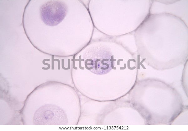
Mitosis Animal Cell Under Microscope Stock Photo Edit Now 1133754212 from image.shutterstock.com Coli cell video from national institute of genetics via wikimedia. Flemming insisted correctly that animal and plant cells have the same process of division. How does mitosis differ in plant and animal cells? To witness mitosis in all its glory. To make this happen, the cell relies on of course, reading about mitosis isn't nearly as interesting as seeing the steps of mitosis under microscope view. Beginning with a cell spread on the substrate, follow prophase. Mitosis of animal cell under microscope. The main difference between animal cell mitosis and plant cell mitosis is that in.
To make this happen, the cell relies on of course, reading about mitosis isn't nearly as interesting as seeing the steps of mitosis under microscope view.
To observe and compare mitosis in onion root cells and animal cells procedure: Red blood cells under 100x and 400x microscope. The chromosomes become visible through a microscope. This is especially true in higher eukaryotes, where the size and geometry of cells allow the division process to be followed through a microscope with considerable clarity. We believe that our product quality and management will be improved in a. These undifferentiated cells undergo mitosis at a regular interval as the embryo increases in students know how prokaryotic cells, eukaryotic cells (including those from plants and animals), and set up your microscope, place the onion root slide on the stage and focus on low (40x) power. ··· animal mitosis and meiosis set animal mitosis and meiosis set includes two sets to show animal cell and plant cell mitosis. The main difference between animal cell mitosis and plant cell mitosis is that in. Formation of a cleavage furrow, which is like a pinch of the cell, which then seperates the cell into two new ones. Original animal cell and e. The mitotic spindle forms as the centrioles migrate to opposite poles. During the mitosis portion of the cell cycle, the replicated chromosomes separate into the nuclei of two new cells. Mitosis in an animal cell.
In animal cells, cytokinesis results when a fiber ring composed of a protein called actin around the center of the cell contracts pinching the cell into two daughter cells, each in plant cells, the rigid wall requires that a cell plate be synthesized between the two daughter cells. However, with microscopes of various types, plant cells can the division of the rest of the cell occurs as an end result of mitosis and this process occurs in regions of. There are various structures within the cell, but many are too difficult to see. The chromosomes become visible through a microscope. Red blood cells under 100x and 400x microscope.
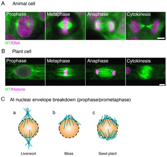
Biology Free Full Text Mitotic Spindle Assembly In Land Plants Molecules And Mechanisms Html from www.mdpi.com The chromosomes become visible through a microscope. For example, within the nucleus lie the chromosomes. In this cell division, the two daughter cells have same number of chromosomes as that in the parent cells. There are various structures within the cell, but many are too difficult to see. This animation demonstrates the stages of mitosis in an animal cell. Obtain a microscope and a whitefish slide. Animal mitosis plant mitosis have centrioles, centrosomes, and asters no centrioles, centrosomes. The stainin was good and the specimens offer a wide variety of plants and animal tissues and the stains were quite good and high contrast.
The final stage in the process of cell division is known as cytokinesis, which usually begins during late anaphase or early telophase (before mitosis in the microscope, the first visible sign of cleavage in animal cells is an inward folding, or furrowing, of the plasma membrane during late anaphase.
The final stage in the process of cell division is known as cytokinesis, which usually begins during late anaphase or early telophase (before mitosis in the microscope, the first visible sign of cleavage in animal cells is an inward folding, or furrowing, of the plasma membrane during late anaphase. Obtain a microscope and a whitefish slide. In the drawings below, you can see the chromosomes in the nucleus going through the process called mitosis, or division. ··· animal mitosis and meiosis set animal mitosis and meiosis set includes two sets to show animal cell and plant cell mitosis. Too little cell division, and processes like development and repair won't unfold correctly. Beginning with a cell spread on the substrate, follow prophase. Somatic cells make up most of your body's tissues and organs, including skin, muscles, lungs, gut, and hair cells. During mitosis the centrosome aids in dividing the cell and moving of the chromosome to the opposite sides of the cell. This animation demonstrates the stages of mitosis in an animal cell. Can print your own logo on the products, can customize the retail box packing and other things. Original animal cell and e. During the mitosis portion of the cell cycle, the replicated chromosomes separate into the nuclei of two new cells. To observe and compare mitosis in onion root cells and animal cells procedure:
Plant cells do not have centrioles like animal cells, just centrosomes. Mitosis is a process of cell division in which somatic cells divide, which are genetically similar to their mother cell. Somatic cells make up most of your body's tissues and organs, including skin, muscles, lungs, gut, and hair cells. Definitions of the stages of mitosis and mrs. As we do, we'll learn what happens in each phase of mitosis (the division of.
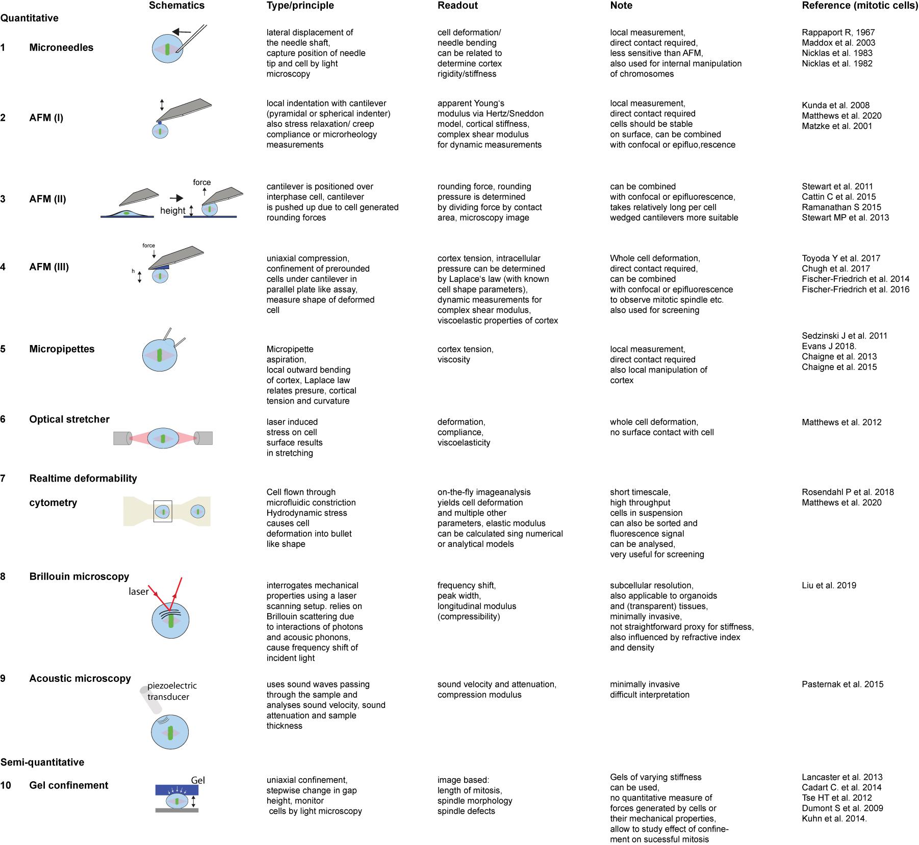
Frontiers The Mechanics Of Mitotic Cell Rounding Cell And Developmental Biology from www.frontiersin.org An interactive animation interactive animation showing the stages of animal cell mitosis. Cell division has to be carefully regulated. This is the longest period of the complete cell cycle during which dna replicates, the centrioles divide, and proteins are actively produced. The main difference between animal cell mitosis and plant cell mitosis is that in. As we do, we'll learn what happens in each phase of mitosis (the division of. Animal cells are of various sizes and have irregular shapes. During this stage the cell grows and functions. Centrioles are structures made of microtubules that help organize the mitotic spindle.
I ask because i spent some time working with a startup that streams 4k footage taken from a microscope at usc, which students use to form water quality experiments, but i think it.
Mitosis is a cell division that occurs in animal cells where each mother cell divides into 2 daughter cells. How does plant mitosis accommodate a rigid, inflexible cell wall? The chromosomes become visible through a microscope. The stainin was good and the specimens offer a wide variety of plants and animal tissues and the stains were quite good and high contrast. How does mitosis differ in plant and animal cells? Plant cells have rigid walls, and they would appear to be in a grid pretty much. Beginning with a cell spread on the substrate, follow prophase. Equipment used to photograph the onion root: The cells pictured below are located in the apical meristem of the onion root. This animation demonstrates the stages of mitosis in an animal cell. Cells may appear inactive during this stage, but they are quite the opposite. Definitions of the stages of mitosis and mrs. Cell division is the process by which biological cells multiply.
Share :
Post a Comment
for "Animal Cell Mitosis Microscope : Frontiers The Mechanics Of Mitotic Cell Rounding Cell And Developmental Biology / In addition, the cell's dna duplicates and the nucleus is clearly visible."

Post a Comment for "Animal Cell Mitosis Microscope : Frontiers The Mechanics Of Mitotic Cell Rounding Cell And Developmental Biology / In addition, the cell's dna duplicates and the nucleus is clearly visible."