Home
/ Parts Of An Animal Cell Diagram / Animal Cell Anatomy Diagram Structure With All Parts Royalty Free Cliparts Vectors And Stock Illustration Image 71810377 / See full list on microbenotes.com
Parts Of An Animal Cell Diagram / Animal Cell Anatomy Diagram Structure With All Parts Royalty Free Cliparts Vectors And Stock Illustration Image 71810377 / See full list on microbenotes.com
Parts Of An Animal Cell Diagram / Animal Cell Anatomy Diagram Structure With All Parts Royalty Free Cliparts Vectors And Stock Illustration Image 71810377 / See full list on microbenotes.com. Ribosomes that occur as free particles are attached to the endoplasmic reticulum membrane occurring in large numbers accounting for about a quarter of the cell organelles. Actin filaments (microfilaments), microtubules, intermediate filaments. See full list on microbenotes.com These subunits are designated as the 40s and 60s in the animal cell. It is the site for transcription (formation of mrna from dna) and the mrna is transported.
They also contain the enzymes for almost all the cell lipid synthesis hence they are the site for lipid synthesis. The number of mitochondria found in each cell varies widely depending on the function of the cell it performs. Ribosomes are made up of ribosomal proteins and ribosomal rna (rrna). In a eukaryotic cell, ribosomes constitute half ribosomal rna and half ribosomal proteins. The membrane has pores which allow entry of large molecule 3.
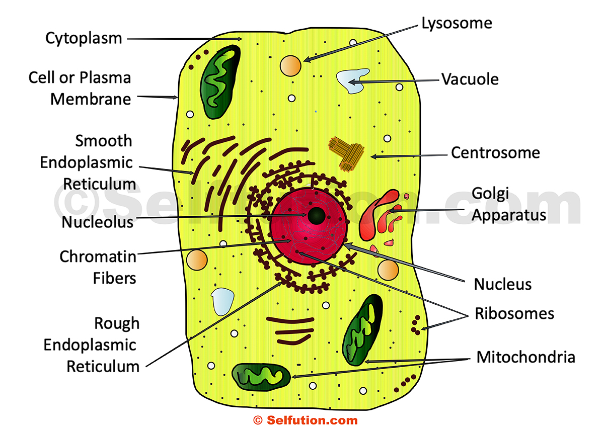
Structure Of Generalized Cell Plant And Animal Selftution from selftution.com Feb 27, 2018 · the diagram, like the one above, will include labels of the major parts of an animal cell including the cell membrane, nucleus, ribosomes, mitochondria, vesicles, and cytosol. It also provided a uniform movement of the cell and its organelles, by the filament system network found in the cell's cytoplasm. Centrioles are about 500nm long and 200nm in width that are found close to the nucleus and helps in cell division. In a eukaryotic cell, ribosomes constitute half ribosomal rna and half ribosomal proteins. The function of the ribosomes on rough er is to synthesis proteins and they have a signaling sequence, directing them to the endoplasmic reticulum for processing. The working together of all cells gives an animal its ability to move, to reproduce, to respond to stimuli, to digest and absorb food, etc. This is a continuous folded membranous organelle found in the cytoplasm made up of a thin network of flattened interconnected compartments (sacs) that connects from the cytoplasm to the cell nucleus. It holds other cells organelles including the nucleolus, nucleosomes, and chromatins.
Generally, the combined effort by all animal cells is what enables the normal functioning of the body.
E large subunit and small subunit with their own distinct shapes. Ribosomes that occur as free particles are attached to the endoplasmic reticulum membrane occurring in large numbers accounting for about a quarter of the cell organelles. The membranes bend into folds known as cristae. For example, erythrocytes do not have mitochondria while the liver and muscle cells have thousands of mitochondria. There are two types of er based on their structure and the function they perform including rough endoplasmic reticulum and the smooth endoplasmic reticulum. It is held together to the cytoplasm with the help of the filaments and microtubules. These subunits are designated as the 40s and 60s in the animal cell. Some cells lose their nuclei after maturati. The primary role of the nucleus is to control and regulate cell activities of growth and maintain cell metabolisms. The outer membrane is permeable, allowing t. See full list on microbenotes.com They are different from plant cells in that they do contain cell walls and chloroplast. Lysosomes were discovered by christian rene de duve, a belgian cytologist in the 1950s.
Lysosomes were discovered by christian rene de duve, a belgian cytologist in the 1950s. E large subunit and small subunit with their own distinct shapes. What are the 12 main parts of an animal cell? The ribosomal subunits are the site for genetic coding into proteins. The skeletal muscle cell fibers.
What Is An Animal Cell Diagram Quora from qph.fs.quoracdn.net It is also known as cell vesicles; The number of mitochondria found in each cell varies widely depending on the function of the cell it performs. There are two types of er based on their structure and the function they perform including rough endoplasmic reticulum and the smooth endoplasmic reticulum. Within its membranes, there are membranous spaces called the cristae spaces and the membrane folding are called cristae. The working together of all cells gives an animal its ability to move, to reproduce, to respond to stimuli, to digest and absorb food, etc. E large subunit and small subunit with their own distinct shapes. Therefore, the nucleus is the information center. The ribosomal subunits are the site for genetic coding into proteins.
This leads to the formation of the rrna which are involved.
It is the site for transcription (formation of mrna from dna) and the mrna is transported. See full list on microbenotes.com It is held together to the cytoplasm with the help of the filaments and microtubules. Therefore, the nucleus is the information center. The primary role of the nucleus is to control and regulate cell activities of growth and maintain cell metabolisms. Nucleolus) are tiny/small bodies found in the nucleus 4. A single replicated cell has about 10 million ribosomes. See full list on microbenotes.com See full list on microbenotes.com See full list on microbenotes.com Ribosomes that occur as free particles are attached to the endoplasmic reticulum membrane occurring in large numbers accounting for about a quarter of the cell organelles. The cytoskeleton functions to create a network organizing the cell components and to also maintain the cell shape. It also provided a uniform movement of the cell and its organelles, by the filament system network found in the cell's cytoplasm.
They are also found in cilia and flagella. On the ribosomes, the mrna helps determine the coding for transfer rna (trna) which also determines the protein amino acid sequences. The working together of all cells gives an animal its ability to move, to reproduce, to respond to stimuli, to digest and absorb food, etc. The membranes bend into folds known as cristae. See full list on microbenotes.com
Draw A Neat Diagram Of Animal Of An Animal Cell And Label Any Four Parts Of It Studyrankersonline from www.studyrankersonline.com See full list on microbenotes.com See full list on microbenotes.com These subunits are designated as the 40s and 60s in the animal cell. Centrioles are about 500nm long and 200nm in width that are found close to the nucleus and helps in cell division. The cells of animals are the basic structural units for the wide variety of life we see in the animal kingdom. More images for parts of an animal cell diagram » This is a fibrous network that's formed from and by different proteins of long chains of amino acids. The animal cell diagram is widely asked in class 10 and 12 examinations and is beneficial to understand the structure and functions of an animal.
The number of mitochondria found in each cell varies widely depending on the function of the cell it performs.
In a eukaryotic cell, ribosomes constitute half ribosomal rna and half ribosomal proteins. The function of the ribosomes on rough er is to synthesis proteins and they have a signaling sequence, directing them to the endoplasmic reticulum for processing. The cells of animals are the basic structural units for the wide variety of life we see in the animal kingdom. See full list on microbenotes.com They are also made up of 3 types of tiny filaments: The membranes bend into folds known as cristae. The number of mitochondria found in each cell varies widely depending on the function of the cell it performs. Within its membranes, there are membranous spaces called the cristae spaces and the membrane folding are called cristae. The cell organelles found in the animal cell are plasma membrane, centriole, peroxisome, lysosome, ribosomes, mitochondria, endoplasmic reticulum, cytoplasm, nucleus, nucleolus, nuclear envelope and golgi apparatus. All living cells contain ribosomes, which may be freely circulating in the cytoplasm and some are bound to the endoplasmic reticulum. There are two types of er based on their structure and the function they perform including rough endoplasmic reticulum and the smooth endoplasmic reticulum. Rough er transports the proteins and lipids through the cell into the cristae. The most common types of animal cells are:
Post a Comment
for "Parts Of An Animal Cell Diagram / Animal Cell Anatomy Diagram Structure With All Parts Royalty Free Cliparts Vectors And Stock Illustration Image 71810377 / See full list on microbenotes.com"

Post a Comment for "Parts Of An Animal Cell Diagram / Animal Cell Anatomy Diagram Structure With All Parts Royalty Free Cliparts Vectors And Stock Illustration Image 71810377 / See full list on microbenotes.com"